
By April Jasper, OD, FAAO, Advanced Eyecare Specialists, West Palm Beach, Florida
Due to an aging population and an increasingly diverse demographic patient base, most optometric practies are seeing more patients who are either at risk for, or already have, glaucoma, the second-leading cause of blindness in the U.S. While there is a lot left to learn about this “silent thief of sight,” there have been groundbreaking studies that have significantly changed the way this disease is treated and managed.
About 20 years ago, the Ocular Hypertension Treatment Study (OHTS) showed that central corneal thickness (CCT) was a significant predictor of which ocular hypertension patients were at higher risk for developing glaucoma.1 While CCT is currently the standard of care in glaucoma decision-making, research has continued, and the focus has shifted from CCT to corneal biomechanics. Today, you can’t pick up a journal on the subject of glaucoma without finding data that shows corneal hysteresis (CH) is significantly more important in glaucoma decision-making.
A 2013 study by Medeiors et al. published in Ophthalmology showed that CH correlates with the rate of visual field progression. Each 1mmHg decrease in CH value was associated with a 0.25 percent per year increase in rate of decline in visual field index (VFI) in a univariable model including only CH as a predictive factor along with time and the interaction (P<0.001). In eyes with a CH of 5mmHg, each 1mmHg increase in IOP was associated with a 0.38 percent per year increase in rate of VFI loss. The results of this study support the idea that CH is an important and independent risk factor for progression in glaucoma patients.2
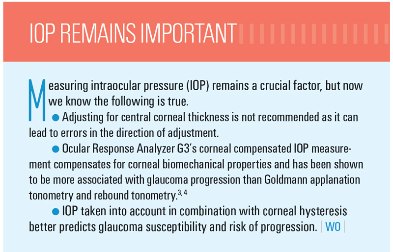
I have been using CH in my practice with the Reichert Ocular Response Analyzer® for years. The measurement is a result of a patented, noncontact, bidirectional applanation process, which is taken with a gentle puff of air on the eye. CH reflects the ability of the cornea to absorb and dissipate energy. In addition, this measurement enables the Ocular Response Analyzer to provide a more accurate intraocular pressure (IOP) reading, called corneal compensated IOP (IOPcc)— closer to true IOP than Goldmann applanation tonometry (GAT).
Like most optometrists, I used to check IOP with GAT and then measured corneal thickness, but I wanted to have a better way to check IOP and have valuable information that I could not obtain otherwise. My primary motivation for using the Ocular Response Analyzer was to predict the rate of progression in my diagnosed glaucoma patients more accurately and also predict their response to therapy (high CH, small IOP reduction/low CH, larger IOP reduction). In addition, it allows me more accurately to predict the risk of my glaucoma suspects. The added information that CH provides me has vastly improved my diagnostic and treatment skills.
Ultimately, I found that in my busy practice, the Ocular Response Analyzer has been a crucial part of my role in preserving my patients’ sight, which is every optometrist’s ultimate goal. I want to make certain that I get the diagnosis and treatment plan right. I also make it a priority to explain to my patients the importance of CH and why it is important. Recently I upgraded my original unit to the latest model, Ocular Response Analyzer G3. All my patients, both young and old, find the test fast, easy, convenient and expected as part of their comprehensive eye health evaluation. Without this invaluable tool and the information it provides, I would not be able to treat my patients with the confidence that I do; it has forever changed my practice for the better.


References:
1 Gordon M, Beiser JA, Brandt JD, Heuer DK, Higginbotham EJ, Johnson CA, Keltner JL, Miller JP, Parrish RK 2nd, Wilson MR, Kass MA. The Ocular Hypertension Treatment Study: baseline factors that predict the onset of primary open-angle glaucoma. Arch Ophthalmol. 2002 Jun;120(6):714- 20; discussion 829-30. 2 Medeiros FA, Meira-Freitas D, Lisboa R, Kuang TM, Zangwill LM, Weinreb RN. Corneal hysteresis as a risk factor for glaucoma progression: a prospective longitudinal study. Ophthalmology. 2013 Aug;120(8):1533-40. 3 Medeiros FA, Weinreb RN. Evaluation of the influence of corneal biomechanical properties on intraocular pressure measurements using the ocular response analyzer. J Glaucoma. 2006 Oct;15(5):364-70. 4 Bianca N. Susanna, Nara G. Ogata, MD, Fábio B. Daga, MD, Carolina N. Susanna, Alberto Diniz-Filho, MD, PhD, Felipe A. Medeiros, MD, PhD. Association between Rates of Visual Field Progression and Intraocular Pressure Measurements Obtained by Different Tonometers. Ophthalmology 10.1016/j.ophtha.2018.07.031


